Back Of Skull Anatomy / Human Skull 3 4 Back View Skull Reference Skull Anatomy Human Skull : Anatomy next provides anatomy learning tools for students and teachers.
Back Of Skull Anatomy / Human Skull 3 4 Back View Skull Reference Skull Anatomy Human Skull : Anatomy next provides anatomy learning tools for students and teachers.. The skull performs vital functions. Anatomy & physiology · anatomy and physiology. Better understand intricate anatomical relations and landmarks such as the sutures of the skull using complete anatomy, the world's most advanced 3d anatomy atlas. Skull contains both junction types: Looking at it from the inside it can be subdivided into.
Synarthrodial joints, which allow no movement. Inferior view of base of the skull. A thorough description is beyond the. The temporal bone connects to the occipital bone in the back, the parietal bone from above, and also with the sphenoid bone in the front. Overview, anterior skull base, middle skull base march 18, 2017.
7 99 Aud 06 Anterior And Posterior Views Of Skull Anatomy Map 14 X25 Poster Ebay Collectibles Anatomy Bones Skull Anatomy Human Anatomy from i.pinimg.com The frontal, parietal, temporal and occipital bones are joined at the cranial sutures. They don't move and united into a single unit. Skull bones aren't fused together at birth. Looking at it from the inside it can be subdivided into. The skull includes the upper jaw and the cranium. Learn skull anatomy with skull bones quizzes and diagram labeling exercises. This anatomic region is complex and poses surgical challenges for otolaryngologists and neurosurgeons alike. The major sutures are the coronal suture, sagittal suture, lambdoid suture and squamosal sutures.
The frontal, parietal, temporal and occipital bones are joined at the cranial sutures.
The frontal, parietal, temporal and occipital bones are joined at the cranial sutures. A cartilaginous mould begins to grow this is why raising your eyebrows can create the appearance that the back of the head is moving. Please feel free to download and print. Anatomy of the skull and bones of cranium on medical illustrations. Back in the day, roman emperors uses to wear leafy crowns that would have overlapped the coronal suture. Understanding the human skull anatomy is necessary for a wide range of professionals from doctors (dentists, oral surgeons, neurosurgeons, etc.) to the structure of the skull bones is to a large extent determined by and interconnected with the anatomy of the sensory organs, situated in the head, as. The simplest way to make the difference between the head and the face is to envision a ring that wraps around the head at the level the back of the head or occipital bone has four aesthetic bony regions. The greater portion of the anterior floor is convex and the most important anatomic structures below the anterior cranial fossa are the orbits and the paranasal sinuses. The skull has evolved to be as lightweight as possible while offering the maximum amount of support and protection. Skull bones aren't fused together at birth. William is a final year medical student in australia who has taught anatomy to tertiary science and. The cranium and the mandible. The bbc is not responsible for the content of external websites.
It offers protection to the brain, eye balls, inner ears, and nasal passages. It supports and protects the face and the brain. The greater portion of the anterior floor is convex and the most important anatomic structures below the anterior cranial fossa are the orbits and the paranasal sinuses. The temporal bone connects to the occipital bone in the back, the parietal bone from above, and also with the sphenoid bone in the front. Anatomy next provides anatomy learning tools for students and teachers.
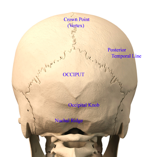
Back Of Head Skull Anatomy Dr Barry Eppley Indianapolis Explore Plastic Surgery from exploreplasticsurgery.com The base of the skull (or skull base) forms the floor of the cranial cavity and separates the brain from the structures of the neck and face. The skull performs vital functions. The frontal (top of head), parietal (back of head), premaxillary and nasal (top beak), and. From an anatomical perspective, the skull is divided into two parts: The major sutures are the coronal suture, sagittal suture, lambdoid suture and squamosal sutures. Skull, skeletal framework of the head of vertebrates, composed of bones or cartilage, which form a unit that protects the brain and some sense organs. Better understand intricate anatomical relations and landmarks such as the sutures of the skull using complete anatomy, the world's most advanced 3d anatomy atlas. They don't move and united into a single unit.
The cranium and the mandible.
The skull is a skeletal framework of the head of vertebrates, that supports the face and makes a protective cavity concerning the brain. Synarthrodial joints, which allow no movement. Anatomy & physiology · anatomy and physiology. The skull base is the inferior portion of the neurocranium. The skull or known as the cranium in the medical world is a bone structure of the head. Looking at it from the inside it can be subdivided into. They don't move and united into a single unit. The skull has a single occipital condyle.7 the skull consists of five major bones: Skull reshaping is done on any of the structures that lie above the face. Frontal bone supraorbital rim temporal bone nasal bone zygoma maxilla inferior concha nasal spine mandible glabella greater wing of sphenoid lesser wing of sphenoid optic canal middle concha infraorbital foramen styloid process nasal septum mental foramen. This is a model of the human (homo sapiens) skull. The base of the skull (or skull base) forms the floor of the cranial cavity and separates the brain from the structures of the neck and face. Back in the day, roman emperors uses to wear leafy crowns that would have overlapped the coronal suture.
Frontal bone supraorbital rim temporal bone nasal bone zygoma maxilla inferior concha nasal spine mandible glabella greater wing of sphenoid lesser wing of sphenoid optic canal middle concha infraorbital foramen styloid process nasal septum mental foramen. Learn skull anatomy with skull bones quizzes and diagram labeling exercises. They don't move and united into a single unit. The skull bones can be classified into two groups: Anatomy & physiology · anatomy and physiology.
I Can Feel A Little Mass Protruding Out When I Touch The Back Of My Skull On Both Sides Of The Hemispheres Is This A Tumor Quora from qph.fs.quoracdn.net Skull, skeletal framework of the head of vertebrates, composed of bones or cartilage, which form a unit that protects the brain and some sense organs. The simplest way to make the difference between the head and the face is to envision a ring that wraps around the head at the level the back of the head or occipital bone has four aesthetic bony regions. Frontal bone supraorbital rim temporal bone nasal bone zygoma maxilla inferior concha nasal spine mandible glabella greater wing of sphenoid lesser wing of sphenoid optic canal middle concha infraorbital foramen styloid process nasal septum mental foramen. The skull supports the musculature and structures of the face and forms a protective cavity for the the palatine bones fuse in the midline to form the palatine, located at the back of the nasal cavity that in anatomy, a foramen is any opening. The skull base is the inferior portion of the neurocranium. A cartilaginous mould begins to grow this is why raising your eyebrows can create the appearance that the back of the head is moving. Inferior view of base of the skull. Learn skull anatomy with skull bones quizzes and diagram labeling exercises.
William is a final year medical student in australia who has taught anatomy to tertiary science and.
The bbc is not responsible for the content of external websites. The skull includes the upper jaw and the cranium. The skull is the bony skeleton of the head. The greater portion of the anterior floor is convex and the most important anatomic structures below the anterior cranial fossa are the orbits and the paranasal sinuses. Anatomy next provides anatomy learning tools for students and teachers. The base of the skull (or skull base) forms the floor of the cranial cavity and separates the brain from the structures of the neck and face. They don't move and united into a single unit. The cranium and mandible was exported from ct data. Foramina inside the body of humans and other animals. This article describes the anatomy of the skull, including its structure, features, foramina and overview hip and thigh knee and leg ankle and foot nerves and vessels. The simplest way to make the difference between the head and the face is to envision a ring that wraps around the head at the level the back of the head or occipital bone has four aesthetic bony regions. William is a final year medical student in australia who has taught anatomy to tertiary science and. It is comprised of many bones, formed by intramembranous ossification, which are joined together by sutures (fibrous joints).


0 Comments